- AdventHealth University

The foundation of modern diagnostic imaging and radiography was established more than 100 years ago when German scientist Wilhelm Conrad Röntgen first observed X-rays in 1895. In 1901, he was the first to be awarded the Nobel Prize in physics for his discovery. Today, much as in all other fields across industries, digitization is transforming the field of radiography.
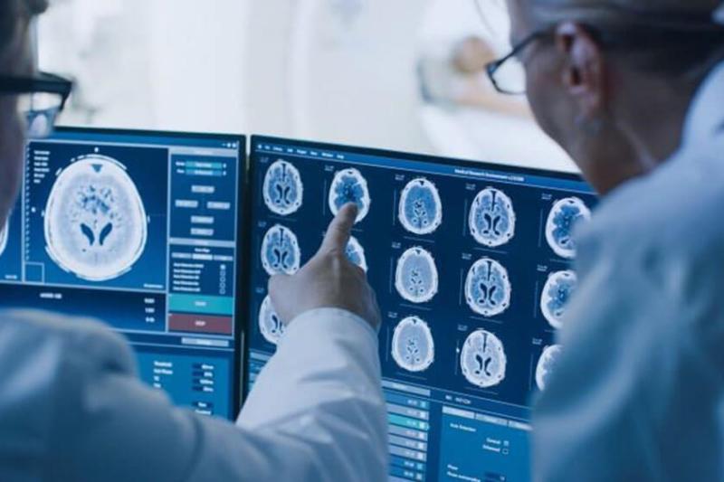
Many advancements have been made in the field of digital radiography in recent years, including AI-aided X-ray interpretation, dual-energy imaging, tomosynthesis, computer-aided diagnosis, automatic image stitching, and digital mobile radiography. These advancements have improved image quality, helping to enhance patient care and support better patient outcomes. Additionally, the use of digital radiography reduces the need for retakes, which provides the benefit of lower radiation exposure.
Digital radiography also offers the following advantages:
- Shorter exposure times
- Improved detail detectability
- Extremely high image quality
- Faster processing and diagnosis
- Images that are easier to store and share with doctors
- Capability for remote viewing from connected digital devices
Imaging professionals looking to advance their careers and participate in the evolving, innovative field of digital radiography can enroll in an online Bachelor of Science in Imaging Sciences program. Pursuing a bachelor’s degree in imaging sciences can help professionals enhance their knowledge of imaging modalities and prepare to move into an advanced radiologic technologist position or lead an imaging department.
Differences Between X-Rays and Digital Radiology
Common functions of both traditional X-rays and digital radiography include image detection, image capturing, and image data storage. During a radiographic procedure, an X-ray beam passes through an individual’s body. Some of the X-rays are absorbed by the internal body structure. The other X-rays are transmitted to a detector, such as film or a digital detector, forming a pattern that is recorded for future evaluation.
The primary differences between traditional X-rays and digital radiography are as follows:
- Traditional X-rays use film to capture images of internal structures in the body.
- In digital radiography, digital detectors instantly produce a digital radiographic image and store the images separately on a digital medium such as a computer.
Digital radiography makes it easy for images to be accessed electronically by radiographers, doctors, and other medical professionals via electronic health records (EHRs) or devices that store patient information.
It can be categorized into two types:
- Computed radiography (CR) works through an indirect process. It replaces traditional film used in traditional radiography with an imaging plate (IP). The information is then transferred over to a computing device for analysis.
- Digital detector array radiography (DDA) is also known simply as digital radiography, which uses a digital detector array or flat panel detector to convert X-rays directly into a digital image.
Digital technology sits at the center of digital radiography. This advanced form of X-ray inspection has become prominent in the medical imaging field over the past decade. New advancements continue to transform the field of radiology. For example, applications such as:
- Virtual reality and augmented reality to create immersive training for future radiology professionals
- Deep mind to reduce false results for cancer screening
- Cardiac MRI segmentation to deliver real-time diagnosis
- Augment intelligence to automate mundane manual tasks
AI-Aided X-Ray Interpretation
With advancements in computer vision, machine learning (ML), artificial intelligence (AI), and deep learning algorithms, the field of radiology has made huge strides in analyzing and interpreting imaging data. Through AI-aided X-ray interpretation, radiology professionals can improve the quality of patient care by speeding up and improving the accuracy of the diagnosis and treatment of injuries and diseases.
AI-aided X-ray interpretation works as follows:
- Using algorithms, it analyzes data and images at high speed from public and proprietary medical databases.
- It then compares images with previous findings to identify patterns and anomalies.
Benefits of AI-aided X-ray interpretation in diagnostic imaging include the following:
- Faster tracking of crucial information for diagnosis
- The ability to prioritize critical cases better
- Reduction of errors in reading electronic health records (EHRs)
Use Cases for AI-Aided X-Ray Interpretation
AI-aided X-ray interpretation has advanced over the last several years, especially regarding chest radiography, which is the most common diagnostic imaging examination in emergency departments. It continues to advance in many areas. Below are a few examples:
- AI-aided X-ray interpretation was used to help diagnose COVID-19. According to a European Radiology study, AI-assisted chest X-ray assessments helped radiologists differentiate COVID-19-positive from COVID-19-negative patients and improved the precision of diagnoses from 65.9% to 81.9%.
- A study published in JAMA found that AI algorithms performed as well as radiology residents when making preliminary interpretations of chest radiographs. The findings of the study suggest that AI can therefore improve radiology workflows, strengthen overall accuracy for more precise diagnoses, help address issues with resource scarcity, and lower healthcare costs.
Dual-Energy Imaging
Dual-energy imaging is a type of digital radiography, specifically computed tomography (CT). CT, also known as computed axial tomography (CAT), works this way:
- It uses a standard X-ray tube that produces X-ray beams that pass through the body at multiple angles and are captured by digital detectors.
- A computer then assembles the captured data to make cross-sectional images of the body that resemble “slices.”
A dual-energy CT scanner works similarly, but it also uses a second, lower-voltage X-ray tube in addition to the standard X-ray tube. The process of using two different X-ray energy sources of varying power provides advantages over standard CT because it produces clearer images for detecting lesions and abnormalities in a faster time.
In some cases, radiology professionals use substances known as contrast agents to aid in imaging. For example, iodine-based contrast is a commonly used agent that allows for examining blood vessels. A dual-energy CT scanner can detect the iodine more clearly than a standard CT scanner, producing more detailed images that can help improve diagnosis.
Use Cases for Dual-Energy Imaging
The concept of dual-energy imaging is not new. Research going as far back as the 1970s and 1980s demonstrated the advantages of dual-energy technology in improving tissue characterization. However, the time required for data acquisition was extensive, limiting dual-energy imaging’s usability for diagnostic imaging.
Today, new technology with faster processors allows for more rapid data acquisition and analysis, increasing the adoption of dual-energy imaging in the medical community.
Dual-energy imaging may be used for the following use cases:
- To produce better images of blood vessels
- To reduce the number of examinations a patient has to undergo
- To detect abnormalities in the body, for example detecting what type of kidney stone is present in a patient
- To improve image quality if a patient has metal inserts in their body structure
- To produce images that are well-designed for advanced 3D reconstruction and visualization of structures
Tomosynthesis
Tomosynthesis, an advanced type of digital mammography approved by the Food and Drug Administration (FDA) in 2011, goes by many names, including 3-D mammography, breast tomosynthesis, and digital breast tomosynthesis (DBT).
Sometimes, the flat images produced by conventional 2D mammography make it difficult for radiologists to detect cancer, including creating areas in the pictures that appear abnormal. This often leads to patients getting called back for additional tests.
Tomosynthesis helps to avoid this using a different imaging approach. It creates multiple images of the breast using both 2D and 3D-like pictures, which are sent to a computer that uses algorithms to assemble the pictures, giving a more complete view of a patient’s breasts.
Use Cases for Tomosynthesis
Tomosynthesis is a type of digital radiography that offers several benefits. Here are some statistics that highlight the advantage of tomosynthesis:
- Johns Hopkins Medicine reports that tomosynthesis can detect 41% more invasive cancers.
- According to Stanford Health Care, tomosynthesis reduces false positives by approximately 15%.
- A study published in European Radiology reveals that radiologists can improve cancer detection and reduce the time to read images with an AI reading support system.
Tomosynthesis can increase accuracy overall, especially when combined with conventional mammography.
Additional benefits for radiology professionals and patients include:
- Detection of breast cancer in the early stages or in patients not showing any symptoms
- Greater accuracy for breast cancer screening for people with dense breasts
- Identification of tumors that traditional mammograms can miss
- Reduction of callbacks and false-positive results
Computer-Aided Diagnosis
Computer-aided diagnosis (CAD) sits at the intersection of medicine and computer science. This form of digital radiography technology is designed to facilitate diagnostic decisions by medical experts.
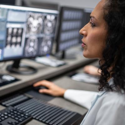
Diagnostic imaging techniques, including X-ray, MRI, CT, and ultrasound diagnostics, provide essential data for radiologists to analyze and evaluate. In most cases, and because of the urgency of finding out the results, radiologists have to read images in a relatively short amount of time.
Types of CAD include computer-aided detection (CADe) and computer-aided diagnosis (CADx).
- Computer-aided detection (CADe): Marks areas of images that appear abnormal to reduce the chances of missing pathologies and to spotlight anomalies.
- Computer-aided diagnosis (CADx): Provides support for assessing and classifying pathologies, such as tumors and lesions, presented in medical images.
Use Cases for Computer-Aided Diagnosis
CAD has been a mainstay in the medical community for many years. The earliest versions of CAD relied on manual engineering and user domain expertise.
Newer approaches to building knowledge in CAD include the use of AI and machine learning. These intelligent systems also process large volumes of complex clinical data to create new knowledge that will enable them to improve diagnostic performance.
CAD software is used in a wide range of medical applications to help radiologists interpret medical images and make more accurate diagnoses. The most common medical applications include breast cancer detection and identification of pulmonary nodules that could cause lung cancer. CAD is also commonly used in the detection and diagnosis of:
- Colon cancer
- Prostate cancer
- Bone metastases
- Coronary artery disease
- Alzheimer’s disease
- Diabetic retinopathy
Automatic Image Stitching
Image stitching in digital radiography, the process of putting together smaller images to form a larger one, is used in various fields. For example, a satellite can take pictures of Earth’s surface, but its range is limited. It can take a picture of the topography of a region in the United States, but not of the whole country. Image stitching leverages intelligent technology to put together the individual images of regions to create a large topographic map of the U.S.
Similarly, in medical applications, a digital radiology scanning system can scan parts of the human body, but not the whole body all at once. If radiology professionals want to see a picture of a patient’s whole bone structure to look for anomalies that are indicative of potential medical issues, they can use image stitching technologies to “stitch” together images of different areas of the body.
Other terms used for image stitching include image registration and image mosaicking. Medical image stitching, by transforming two or more sets of imaging data into one coordinate system, plays a central role in medical robotics and intelligent systems from diagnostics and surgical planning to real-time guidance and post-procedural assessment.
Automatic image stitching is the process of stitching together multiple images without having to have a human intervene in the process. Using AI and ML algorithms, automatic image stitching overlaps regions of the same area to create a panoramic image that enables doctors to diagnose a disease in cases where a single scan does not provide an entire view of the area being investigated.
Use Cases for Automatic Image Stitching
Automatic image stitching can be applied medically in various ways. For example, the stitching of images can allow surgeons to do preoperative planning for surgeries such as leg alignment operations. Additionally, health professionals can see an entire image of a patient’s spine to diagnose scoliosis.
Recent studies offering insight into use cases for automatic image stitching include:
- A recent article in the Sensors journal published on MDPI points out how a panoramic stitching algorithm used in cone beam computed tomography reduced the need for patients to reposition themselves when images are being taken.
- An article in Scientific Reports notes that image stitching can be used to bring together different images of a single area for comparison purposes. For example, scans taken at different times can help to determine the growth of a tumor or determine the effects of surgery.
Digital Mobile Radiography
Digital mobile radiography equipment has wheels and is designed for easy transport in healthcare settings, such as hospitals, where it can be moved to a patient’s bedside. For example, patients with limited mobility following surgery can get imaged by a mobile X-ray unit to help the surgeon determine the outcome of the surgery. The ease of mobility of digital mobile radiography equipment makes it simpler to disinfect.
Digital mobile radiography equipment is a critical tool in digital radiography to help doctors assess and determine the condition of patients before sending them off to more advanced imaging techniques. For example, during the COVID-19 pandemic, hospitals have experienced high volumes of patients who needed diagnostic imaging. Digital mobile radiography units have been used as an essential triaging and screening tool for assessing the lung conditions of patients.
Use Cases for Digital Mobile Radiography
Digital mobile radiography equipment has many clinical diagnostic applications. Here are a few examples.
- Digital mobile radiography equipment allows patients who cannot be moved to a radiology department to get chest radiography.
- Because of its unique size, digital mobile radiography equipment can be positioned within tight spaces and between beds and other equipment, such as in surgical or endoscopic suites.
- According to a study published in BMC Health Services Research, digital mobile radiography equipment can be used outside of the hospital, including in nursing homes for the elderly, group dwellings for people with intellectual disabilities, and shelters.
How to Become a Radiologic Technologist
For individuals interested in advancing their careers as radiologic technologists, it’s important to understand that the work environment includes regularly working with equipment that uses radiation. However, radiation hazards are minimized through the use of personal protective equipment, barriers that protect technologists from exposure, and best practices.
Education and Training
Radiologic technologists are expected to have a college degree from a program accredited by the Joint Review Committee on Education in Radiologic Technology (JRCERT). The minimum requirement is typically an associate degree, while those pursuing advanced positions may need a bachelor’s degree. To become licensed, they need to pass a national certification exam from a credentialing organization such as the American Registry of Radiologic Technologists (ARRT), with some states requiring a specific state certification exam. Certification for MRI technologists is available from the ARRT as a post-primary certification or as a primary certification from the American Registry of Magnetic Resonance Imaging Technologists (ARMRIT).
Important Skills
Important skills in the digital radiography field include detail orientation and technical skills to operate complex equipment, math skills to properly calculate radiation doses, the ability to stand on one’s feet and help patients move and position themselves properly, and interpersonal skills to put patients at ease and collaborate with doctors.
Salary and Job Outlook
According to the U.S. Bureau of Labor Statistics, the median annual salary for radiologic technologists is $61,900 as of May 2020. For magnetic resonance imaging technologists, the median annual salary is $74,690 as of May 2020. The radiologic technologist field is expected to grow by 9% from 2020 to 2030.
Advance Your Career in Digital Radiography
Keeping up with new developments in the field of digital radiography is important if your aim is to move into a high-level technologist position or lead a radiology department. If you are an imaging professional looking to get ahead, a bachelor’s degree in imaging sciences can help you build the expertise to understand the necessary technology and keep up with its advancements.
AdventHealth University Online’s Bachelor of Science in Imaging Sciences allows imaging professionals to advance their knowledge in imaging technology and learn skills including management, marketing, finance, organizational behavior, and business. Students can also customize their education to achieve their career goals, choosing from six specialty tracks: Imaging Leadership, CT, MRI, Vascular Interventional, Sonography, and Interdisciplinary. Explore the curriculum and discover how you can move your imaging career forward at AdventHealth University Online.
Recommended Readings
MRA vs. MRI: What Are the Differences and Uses?
What Is Vascular Interventional Radiology?
Sources:
Advanced Intelligent Systems, “Image Registration in Medical Robotics and Intelligent Systems: Fundamentals and Applications”
American Registry of Magnetic Resonance Imaging Technologists, Join the American Registry of Magnetic Resonance Imaging Technologists
American Registry of Radiologic Technologists, What Is ARRT Certification and Registration?
BMC Health Services Research, “Mobile X-Ray Outside the Hospital: A Scoping Review”
Canadian Association of Radiologists Journal, “Artificial Intelligence Solutions for Analysis of X-Ray Images”
Element, “The Difference Between Computed Radiography (CR) and Digital Radiography (DR)”
European Radiology, “Artificial Intelligence-Assisted Chest X-Ray Assessment Scheme for COVID-19”
Expert Systems with Applications, “A Systematic Survey of Computer-Aided Diagnosis in Medicine: Past and Present Developments”
Healthline, “What You Need to Know About Tomosynthesis for Breast Cancer”
History, “German Scientist Discovers X-Rays”
Inside Radiology, Dual Energy CT Scan
ISRRT, “International Covid-19 Support for Radiographers and Radiological Technologists”
JAMA, “Comparison of Chest Radiograph Interpretations by Artificial Intelligence Algorithm vs Radiology Residents”
Johns Hopkins Medicine, 3-D Mammography
Journal of Physics Conference Series, “Image Stitching for Chest Digital Radiography Using the SIFT and SURF Feature Extraction by RANSAC Algorithm”
The Nobel Prize, Wilhelm Conrad Röntgen
Radiopaedia, Computer Aided Diagnosis
Radiopaedia, Dual Energy CT
Scientific Reports, “A Deep Learning Based Framework for the Registration of Three Dimensional Multi-Modal Medical Images of the Head”
Sensors, “Research on Panoramic Stitching Algorithm of Lateral Cranial Sequence Images in Dental Multifunctional Cone Beam Computed Tomography”
Stanford Health Care, Tomosynthesis (3D Mammography)
TWI, “What Is Digital Radiography and How Does It Work?”
U.S. Bureau of Labor Statistics, Radiologic and MRI Technologists
U.S. Food and Drug Administration, Computed Tomography (CT)
U.S. Food and Drug Administration, Radiography
Windsor Imaging, “The History of the Digital X-Ray”
World Health Organization, “Portable Digital Radiography System: Technical Specifications”


